Gastroenterology
 Gastroenterology
 Ten years of GI imaging science, applied to clinical trials. From structural MRI to motility assessment, we bring a depth of GI expertise that few imaging CROs can match.

Why GI imaging demands a different approach.
The gastrointestinal tract is a dynamic organ system and imaging it well in a clinical trial context is genuinely difficult. Disease activity fluctuates, histological endpoints correlate poorly with imaging findings, and standard scoring systems like MaRIA and sMARIA were developed for research studies, not drug development. Capturing the information that actually matters for a go / no-go decision requires a scientific partner who understands both the biology and the limitations of current methodology and can design endpoints that go further.
Where we have experience.
MRI-based assessment of luminal and transmural disease activity, including bowel wall thickness, enhancement and motility.
Imaging support for UC trials including MRI, colonoscopy and augmented scoring beyond standard endoscopy-derived endpoints.
Proprietary MRI-based motility analysis capturing peristalsis, wall movement and functional bowel and stomach behaviour endpoints unavailable through conventional methods.
Structural and functional imaging support for mucosal healing endpoints in celiac disease trials, including small bowel assessment.
Imaging characterisation of bowel function and motility for functional GI disorders, where structural imaging alone is insufficient.
Imaging endpoint support for early-phase colorectal and gastric programs, including response assessment and structural characterisation.
We work across the full range of GI and hepatic indications. If your area is not listed, speak to our scientific team about your program.

Fidēs was built from a decade of GI imaging research at Motilent. Our approach is not to replicate clinical practice in a trial setting, it is to design imaging endpoints that answer the biological questions your study needs to resolve.
Augmented endpoints beyond MaRIA
Standard MRI scoring systems for IBD MaRIA, sMARIA, the Nancy Index were designed for research, not drug development. They compress biologically meaningful variation into categorical scores, reducing sensitivity to the kinds of early change a Phase II study needs to detect.
Fidēs advises on and implements augmented endpoint approaches that preserve quantitative signal: wall thickness, T2 relaxometry, dynamic contrast enhancement. Providing richer, more sensitive measures of treatment response alongside standard read-outs.

Motility and movement as an endpoint
GI function is not static. Bowel wall movement, peristaltic frequency and motility patterns are biologically meaningful signals in IBD and motility disorders that standard structural MRI does not capture.
Drawing on Motilent’s proprietary motion analysis tools, Fidēs can quantify bowel and stomach motility from cine-MRI sequences. Turning a qualitative radiological observation into a reproducible, protocol-defined quantified endpoint. This is a capability not available through conventional imaging CROs.
Imaging integrated into go / no-go decisions
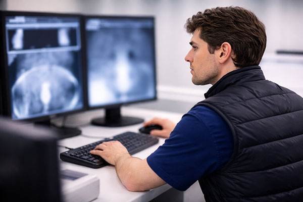
Too often, imaging data arrives as a reporting deliverable at study close: useful for the archive, but too late to influence the decisions that matter. In early-phase development, imaging should be embedded into the scientific thinking from protocol design onwards.
Fidēs works with sponsors from study design through to final analysis to ensure imaging endpoints are aligned with the biological questions that will drive the go / no-go decision and that data quality and interpretability are maintained throughout, not assessed retrospectively.
Real-time visibility matters in GI.
Disease activity in GI trials fluctuates. Sites vary in protocol adherence. Waiting for batch reads at study milestones means weeks of data quality problems that have already compounded by the time they surface. Aperis gives sponsors and study teams live access to imaging data, annotations and site performance throughout the trial so issues are caught early and the scientific integrity of the study is maintained from first scan to last.
- Real-time data visibility Sponsors access imaging data, annotations and analyses continuously, not only at formal reporting milestones.
- Collaborative review environment Multi-modality side-by-side image review with embedded workflows and direct sponsor access.
- Rapid study setup Standard studies configured within one day, complex studies within days, not weeks.
- Adaptive imaging support Designed for live studies where strategy may need to evolve as data emerges.

GI in practice.
 Gastroenterology
 The Role of Movement in Characterising Treatment Response in IBD
Augmenting standard MRI endpoints with motility measures to deliver a more complete characterisation of biological response in Crohn's disease.
Read case study
Gastroenterology The Power of Movement: Motion as an Imaging Endpoint
Augmenting standard MRI endpoints with motility measures to deliver a more complete characterisation of biological response in Crohn's disease.
Read case study
Gastroenterology Structural and Functional MRI Assessment in Post-Surgical J-Pouch Patients
Combining structural and functional MRI to characterise pouch function and inflammation in post-surgical J-pouch patients.
Read case study
Gastroenterology Augmented MRI Endpoints in a Phase II Crohn’s Program
Adding functional motility metrics to a Phase II Crohn's imaging protocol to sharpen the read on treatment response.
Read case study
Gastroenterology Cine-MRI Motility Analysis as a Primary Endpoint
Using cine-MRI motility analysis as a primary endpoint to detect functional change earlier than structural measures alone.
Read case study